Circadian rhythm and circadian sensitivity
 11 апреля, 2014
  admin
admin The human wake-sleep rhythm has a period of approximately 24 hours and the rhythm therefore is referred to as the circadian rhythm or circadian cycle, with the name being derived from the Latin words circa and dies (and its declination diem), meaning approximately and day, respectively. Light has been known for a long time to be the synchronizing clock (zeitgeber) of the human circadian rhythm. For reviews on the development of the understanding of the circadian rhythm including the identification of light as the dominant trigger for the endogenous zeitgeber, see Pittendrigh (1993) and Sehgal (2004).
The wake-sleep rhythm of humans is synchronized by the intensity and spectral composition of light. Sunlight is the natural zeitgeber. During mid-day hours sunlight has high intensity, a high color temperature, and a high content of blue light. During evening hours, intensity, color temperature, and blue content of sunlight strongly decrease. Humans have adapted to this variation and the circadian rhythm is most likely synchronized by the following three factors: intensity, color temperature, and blue content.
Exposure to inappropriately high intensities of light in the late afternoon or evening can upset the regular wake-seep rhythm and lead to sleeplessness and even serious illnesses such as cancer (Brainard et al., 2001; Blask et al, 2003). It is therefore highly advisable to limit exposure to high intensity light in the late afternoon and evening hours, to not be counterproductive to the natural circadian rhythm (Schubert, 1997).
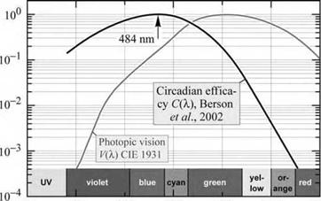
It was believed for a long time that rod cells and the three types of cone cells are the only optically sensitive cells in the human eye. However, Brainard et al. (2001) postulated that an unknown photoreceptor in the human eye would control the circadian rhythm. Evidence presented by Berson et al. (2002) and Hattar et al., (2002) indicates that retinal ganglion cells have an optical sensitivity as well. For a schematic illustration of ganglion cells, see Fig. 16.1. The spectral sensitivity of mammalian ganglion cells was measured and the responsivity curve is shown in Fig. 16.9. Inspection of the figure reveals a ganglion-cell peak-sensitivity at 484 nm, i. e. in the blue spectral range.
Berson et al. (2002) presented evidence that the photosensitive ganglion cells are instrumental in the control of the circadian rhythm. Due to their sensitivity in the blue spectral range, it can be hypothesized that the blue sky occurring near mid-day is a strong factor in synchronizing the endogenous circadian rhythm. The photosensitive ganglion cells have therefore been referred to as blue-sky receptors.
|
|
|
Fig. 16.9. Circadian efficacy curve derived from retinal ganglion cell photoresponse measurements. The ganglion cells on which the measurements were performed originated from mammals. The figure reveals the significant difference between circadian and visual sensitivity (adapted from Berson et al., 2002). |
|
<< U CZ CJ £ і» |
|
-o — u |
|
350 |
|
400 |
|
450 500 550 Wavelength A, (nm) |
|
600 |
|
650 |
Inspection of the spectral sensitivity of the ganglion cells shown in Fig. 16.9 reveals the huge difference of red light and blue light for circadian efficacy: The efficacy of blue light in synchronizing the circadian rhythm can be three orders of magnitude greater than the efficacy of red light. This particular role of blue light should be taken into account in lighting design and the use of artificial lighting by consumers.


 Опубликовано в